Free PDF 2026 Perfect SPI: Real Sonography Principles and Instrumentation Testing Environment
Wiki Article
BTW, DOWNLOAD part of TestPassKing SPI dumps from Cloud Storage: https://drive.google.com/open?id=1cdBFAn3E6c8fKokLelE9lhH3w4BFmZLi
Decades of painstaking efforts have put us in the leading position of SPI training materials compiling market, and the excellent quality of our SPI guide torrent and high class operation system in our company have won the common recognition from many international customers for us. With the high class operation system, we can assure you that you can start to prepare for the SPI Exam with our study materials only 5 to 10 minutes after payment since our advanced operation system will send the SPI exam torrent to your email address automatically as soon as possible after payment.
I know that all your considerations are in order to finally pass the SPI exam. Our SPI study materials have helped many people pass the exam and is about to help you. The 99% pass rate of our SPI training prep is enough to make you feel at ease. Of course, we do everything we could do to ensure that you could think through it and that you also needed to pay a bit of your effort. And with our SPI Exam Questions, you will pass the exam for sure.
>> Real SPI Testing Environment <<
My Review On ARDMS SPI Exam Questions
When dealing with any kind of exams, the most important thing is to find a scientific way to review effectively. Our SPI practice materials compiled by the most professional experts. Till now, we have over tens of thousands of customers around the world supporting our SPI exam torrent. If you are unfamiliar with our SPI Study Materials, please download the free demos for your reference. To some unlearned exam candidates, you can master necessities by our SPI practice materials quickly So our materials are elemental materials you cannot miss.
ARDMS SPI Exam Syllabus Topics:
| Topic | Details |
|---|---|
| Topic 1 |
|
| Topic 2 |
|
| Topic 3 |
|
| Topic 4 |
|
| Topic 5 |
|
ARDMS Sonography Principles and Instrumentation Sample Questions (Q23-Q28):
NEW QUESTION # 23
Which component of a contrast agent causes a marked mismatch in impedance between the agent and blood?
- A. Viscous liquid
- B. Serous liquid
- C. Gas
- D. Solid
Answer: C
Explanation:
Contrast agents used in ultrasound imaging typically consist of microbubbles filled with gas. The significant mismatch in acoustic impedance between the gas in the microbubbles and the surrounding blood creates strong reflectors of the ultrasound waves, enhancing the echogenicity of blood and improving the visibility of blood flow and tissue perfusion. The high contrast provided by the gas-filled microbubbles makes them particularly effective as ultrasound contrast agents.
Reference:
ARDMS Sonography Principles & Instrumentation Guidelines
Kremkau FW. Sonography Principles and Instruments. 9th ed. Philadelphia, PA: Elsevier; 2016.
NEW QUESTION # 24
How is intensity of an ultrasound beam measured?
- A. Hydrophone
- B. Doppler equation
- C. Reynold's number
- D. Autocorrelation
Answer: A
Explanation:
The intensity of an ultrasound beam is measured using a hydrophone. A hydrophone is a specialized device that detects and measures the acoustic pressure of the ultrasound waves in water or tissue-mimicking materials. It is highly sensitive and can measure the variations in pressure, which are used to calculate the intensity and other acoustic parameters of the ultrasound beam.
Reference:
ARDMS Sonography Principles and Instrumentation guidelines
Hoskins, P. R., Thrush, A., Martin, K., & Whittingham, T. A. (2010). Diagnostic Ultrasound: Physics and Equipment.
NEW QUESTION # 25
In this image, what does the data below the baseline represent?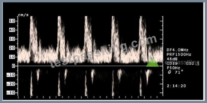
- A. Aliasing and retrograde blood flow
- B. Blood flow directed towards the transducer
- C. Wall filter setting too high
- D. Mirror image artifact
Answer: A
Explanation:
In the provided image, data below the baseline represents blood flow moving away from the transducer, which can indicate retrograde flow. When using spectral Doppler, the baseline separates flows towards and away from the transducer. Aliasing occurs when the velocity of blood flow exceeds the Nyquist limit, causing the display to wrap around and appear on the opposite side of the baseline. This phenomenon is common in high-velocity flow situations and results in part of the flow being displayed below the baseline. Retrograde flow further supports this, as it shows blood moving in the opposite direction to the expected flow.
Reference:
ARDMS Sonography Principles & Instrumentation Guidelines
Kremkau FW. Sonography Principles and Instruments. 9th ed. Philadelphia, PA: Elsevier; 2016.
NEW QUESTION # 26
Which is a method to reduce noise?
- A. Increase frequency
- B. Decrease depth
- C. Decrease beam width
- D. Increase persistence
Answer: D
Explanation:
Comprehensive and Detailed Explanation From Exact Extract:
Persistence averages multiple frames to smooth out random noise fluctuations, reducing the appearance of noise in the image and improving image quality.
According to sonography instrumentation reference:
"Persistence reduces noise by averaging data from consecutive frames, improving signal-to-noise ratio and producing a smoother image." Therefore, the correct answer is D: Increase persistence.
-
NEW QUESTION # 27
A Doppler shift is 10,000 Hz at an angle of flow of 60 degrees. What is the Doppler shift at 0 degrees?
- A. 10,000 Hz
- B. 20,000 Hz
- C. 5,000 Hz
- D. 2,500 Hz
Answer: B
Explanation:
depends on the angle between the ultrasound beam and the direction of blood flow. The Doppler equation includes a cosine function of the angle of insonation (#). At 60 degrees, the cosine is 0.5, and at 0 degrees (parallel to the flow), the cosine is 1. Thus, if the Doppler shift is 10,000 Hz at 60 degrees, it would double to
20,000 Hz at 0 degrees because the cosine of 0 degrees is 1 (cos(0°) = 1) and the cosine of 60 degrees is 0.5 (cos(60°) = 0.5). The formula is: Doppler shift at 0 degrees = Doppler shift at 60 degrees / cos(60 degrees) =
10,000 Hz / 0.5 = 20,000 Hz.
References:ARDMS Sonography Principles and Instrumentation (SPI) Review, Doppler Shift and Angle of Insonation section.
NEW QUESTION # 28
......
We are popular not only because we own the special and well-designed SPI exam materials but also for we can provide you with well-rounded services beyond your imagination. At the very beginning, we have an authoritative production team and our SPI study guide is revised by hundreds of experts, which means that you can receive a tailor-made SPI Study Material according to the changes in the syllabus and the latest development in theory and breakthroughs. Without doubt, our SPI practice torrent keep up with the latest information.
Interactive SPI EBook: https://www.testpassking.com/SPI-exam-testking-pass.html
- Study Materials SPI Review ???? SPI Sample Test Online ???? SPI Latest Material ???? Download ➥ SPI ???? for free by simply searching on ⮆ www.torrentvce.com ⮄ ????Best SPI Practice
- Avail Newest Real SPI Testing Environment to Pass SPI on the First Attempt ???? Simply search for { SPI } for free download on ➠ www.pdfvce.com ???? ????Valid SPI Test Camp
- Top Real SPI Testing Environment | High-quality Interactive SPI EBook: Sonography Principles and Instrumentation 100% Pass ???? Enter ➠ www.dumpsquestion.com ???? and search for ➠ SPI ???? to download for free ????Demo SPI Test
- SPI Sample Test Online ???? SPI Reliable Test Objectives ???? SPI Reliable Test Objectives ???? Search on “ www.pdfvce.com ” for ➽ SPI ???? to obtain exam materials for free download ????Test SPI Guide Online
- Pass Guaranteed Latest ARDMS - SPI - Real Sonography Principles and Instrumentation Testing Environment ???? Enter ➽ www.torrentvce.com ???? and search for ➤ SPI ⮘ to download for free ????Demo SPI Test
- Latest SPI Exam Guide ???? Valid SPI Test Voucher ???? Valid SPI Test Camp ???? Copy URL { www.pdfvce.com } open and search for [ SPI ] to download for free ????Best SPI Practice
- Pass Guaranteed Latest ARDMS - SPI - Real Sonography Principles and Instrumentation Testing Environment ???? Search on ➽ www.vce4dumps.com ???? for ⮆ SPI ⮄ to obtain exam materials for free download ????Best SPI Practice
- Sonography Principles and Instrumentation latest study material - SPI valid vce exam - Sonography Principles and Instrumentation pdf vce demo ⬆ Enter 《 www.pdfvce.com 》 and search for ➡ SPI ️⬅️ to download for free ????SPI Sample Test Online
- New SPI Exam Fee ???? New Study SPI Questions ???? Latest SPI Exam Guide ???? Search for ➡ SPI ️⬅️ on ➤ www.dumpsmaterials.com ⮘ immediately to obtain a free download ????SPI Lead2pass
- Real SPI Testing Environment Pass-Sure Questions Pool Only at Pdfvce ???? Go to website ➤ www.pdfvce.com ⮘ open and search for [ SPI ] to download for free ????New Study SPI Questions
- Top Real SPI Testing Environment | High-quality Interactive SPI EBook: Sonography Principles and Instrumentation 100% Pass ???? Open 《 www.pdfdumps.com 》 enter ⮆ SPI ⮄ and obtain a free download ????SPI Reliable Real Test
- baidubookmark.com, binksites.com, ronaldotqc505629.blogdemls.com, lewisbhgk962989.blog-ezine.com, socialtechnet.com, larajnpp861345.glifeblog.com, rafaelwcie800914.yomoblog.com, mysocialport.com, links2directory.com, esmeeigpz485278.cosmicwiki.com, Disposable vapes
What's more, part of that TestPassKing SPI dumps now are free: https://drive.google.com/open?id=1cdBFAn3E6c8fKokLelE9lhH3w4BFmZLi
Report this wiki page